
Figure EV5. Cho2 expression affects Atg8–PMME formation and mitophagy.
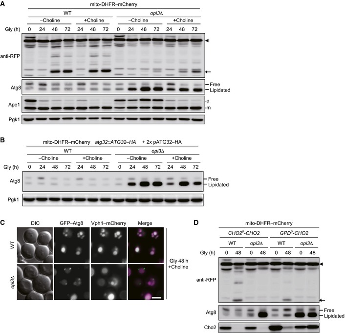
- Choline supplementation partially rescues mitophagy in cells lacking Opi3. Wild‐type and opi3Δ cells expressing mito‐DHFR–mCherry (depicted by arrowhead) cultured in the presence or absence of choline were harvested at the indicated time points and subjected to Western blotting. Generation of free mCherry (depicted by arrow) indicated transport of mitochondria to the vacuole. Pgk1 was monitored as a loading control.
- Atg8 lipidation is partially reduced by choline treatment in opi3‐null cells overexpressing Atg32. Wild‐type and opi3Δ cells expressing mito‐DHFR–mCherry and a chromosomally encoded Atg32–HA were transformed with two Atg32–HA‐expressing plasmids, cultured in the presence or absence of choline, and analyzed as in Fig 7A. Pgk1 was monitored as a loading control.
- Atg8 does not form aggregates in the opi3‐null mutant supplemented with choline. Wild‐type and opi3Δ cells expressing chromosomally encoded Vph1–mCherry (a vacuolar marker) and GFP–Atg8 were pre‐grown in dextrose medium, incubated in glycerol medium (Gly) for 48 h in the presence of choline, and observed using fluorescence microscopy. Scale bar, 2 μm.
- Overexpression of Cho2 leads to partial suppression of mitophagy in wild‐type cells. Wild‐type and opi3Δ cells expressing mito‐DHFR–mCherry (depicted by arrowhead) and Cho2 under the endogenous (CHO2 p ‐CHO2) or strong constitutive (GPD p ‐CHO2) promoter were grown in glycerol medium (Gly) for 48 h, harvested, and subjected to Western blotting. Generation of free mCherry (depicted by arrow) indicated transport of mitochondria to the vacuole.