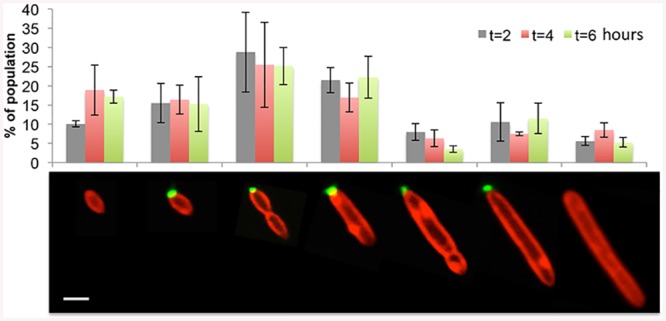
Fig 3. Cell type distribution does not vary with culture age.

Seven different cell types were characterized by size, labeling with Alexa 488-conjugated WGA (green) and presence of a septum using membrane stain FM4-64 (red). Each cell type was quantified at 2, 4 and 6 hours of a culture in early growth stages; n > 300 cells. Scale bar corresponds to 1 μm.
