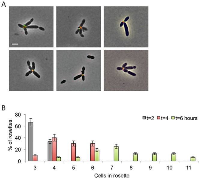
Fig 4. Kinetics of rosette formation.

(A) Rosettes form by cell encounters. Two independent aliquots from the same synchronized culture were stained with either Alexa 488-conjugated WGA (green) or Alexa 594-conjugated WGA (red) and were mixed for 30 minutes at room temperature in buffer prior to imaging. Rosettes with dual-labeled centers are shown. Images are overlay of phase contrast (gray) with green and red fluorescence channels. (B) Rosette complexity increases over time. The number of cells per rosette was quantified over time. Error bars indicate standard deviation of two biological replicates; n > 300 cells.
