
Fig 6. Self-DNA prevents attachment to an abiotic surface.
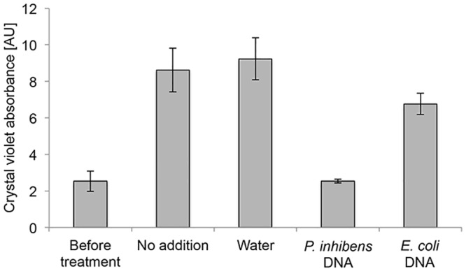
Crystal violet absorbance of attached cells was quantified before and after addition of genomic DNA originating from either P. inhibens or E. coli. “Before treatment” measurement was conducted after 2 hours of incubation in an attachment assay (see Methods) and the different treatments were then applied. Measurements after treatments were conducted one hour following the addition of water or DNA. Error bars indicate standard deviation of at least two biological replicates. Note: crystal violet absorbance values before treatment and after treatment should be compared to time points 2 and 3 hours, respectively, in Fig 5A.