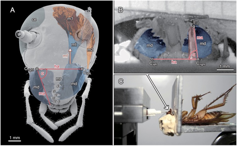
Fig 1. Morphology of the mandibular apparatus of P. americana, kinematics and general experimental setup.
A) Morphology of the cockroach head from a μCT scan with emphasis to the mandibles (coloured light blue) and their driving muscles (light red). 1l-4l: teeth of the left mandible; 1r-3r: teeth of the right mandible; α: opening angle of the right mandible (approx.. 70°); ‘ce’: right complex eye; ‘md’: mandibles; ‘Cleft‘ and ‘Cright’: anterior condyles of the left and right mandible joints; ‘m1’: left mandible closer muscle; ‘m2’: left mandible opener; mf: main direction of the muscle force; ‘mt’: apodeme connecting the mandible closer muscle with the median edge of the mandible; mr: molar region B) Camera view onto the mandibles (light blue) and the sensor tip (light red) during the bite experiments. The horizontal line is defined by the anterior condyles of the left and right mandible joints Cleft and Cright; ‘1r’ depicts the distal tip of the right mandible and α is the mandible angle with respect to the horizontal line, i.e. the opening angle of the right mandible before mathematical correction (see methods). C) Side view of the general setup with the fixated specimen at the right and the force transducer at the left side.

