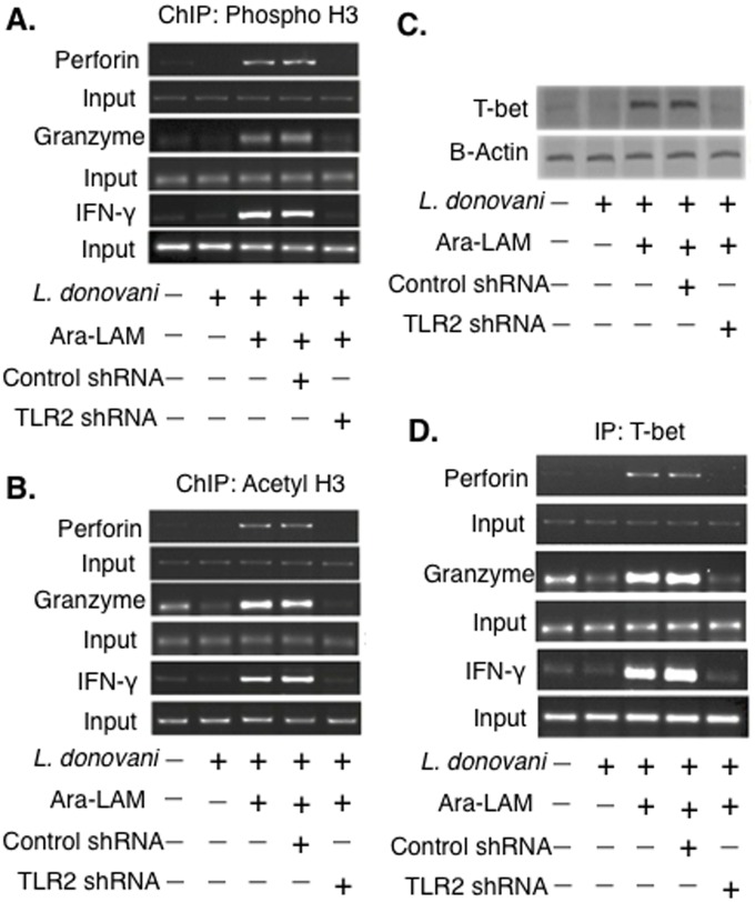
Fig 3. Histone H3 modifications at the IFN-γ, Perforin, Granzyme-B promoter of CD8+ T-cells in different groups of BALB/c mice.
(A-B) CD8+ T cells from differently treated mice groups were co-cultured with autologous infected macrophages for 45 min and chromatin immunoprecipitation (ChIP) assays were conducted. Immunoprecipitations were performed using Abs specific to phosphorylated H3 histone (IP: phospho-H3), acetylated H3 histone (IP: acetyl-H3) and conventional RT PCR was performed using primers specific to the IFN-γ, perforin and granzyme-B promoter region. (C) CD8+ T-cells were co-cultured with infected macrophages, lysed and the nuclear protein extracts were analyzed for the activation of T-bet by Western blot. (D) CD8+ T-cells from differently treated mice group were co-cultured with autologous L. donovani–infected macrophages, Immunoprecipitations were conducted using T-bet (IP: T-bet) specific Abs. Conventional RT-PCR was performed for amplifying the putative T-bet binding sites of the IFN-γ, perforin, granzyme-B promoter. Data represented were one of the three indepenedent experiments with similar results performed in the same way.

