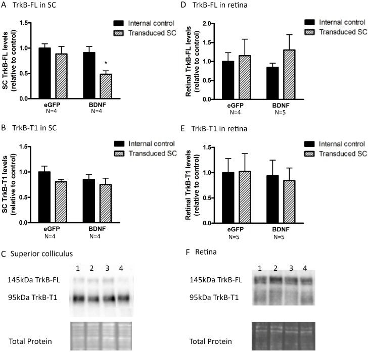
Fig 8. Collicular and retinal TrkB levels upon unilateral viral vector delivery to the SC.
(A) Western blot analysis shows reduced TrkB-FL levels in BDNF vector-transduced SC compared to both internal controls and eGFP vector-transduced animals. (B) Collicular TrkB-T1 levels are not influenced by unilateral SC vector transduction. (C) Western blot for TrkB-FL and TrkB-T1 in the SC 18 d after viral vector delivery showing representative bands for the graphs in A and B, with 1) internal control SC of eGFP vector injected mice, 2) SC transduced with eGFP vector, 3) internal control SC of BDNF vector-transduced mice, 4) BDNF vector-injected SC. (D, E) Upon unilateral SC transduction with eGFP or BDNF vector, no differences in full-length (D) or truncated (E) forms of TrkB are detected in retinas ipsilateral or contralateral to the transduced SC. F. TrkB-FL and TrkB-T1 western blot detection in the retina 18 d after viral vector delivery, representative bands for the graphs in C and D, with 1) internal control retina ipsilateral to eGFP vector-injected SC, 2) retina contralateral to the SC transduced with eGFP vector, 3) internal control retina ipsilateral to BDNF vector-transduced SC, 4) retina contralateral to BDNF vector-injected SC. Key: SC, superior colliculus; BDNF, brain-derived neurotrophic factor; TrkB, tropomyosin receptor kinase B; TrkB-FL, TrkB full-length form; TrkB-T1, TrkB truncated form 1, eGFP, enhanced green fluorescent protein.

