Figure 5.
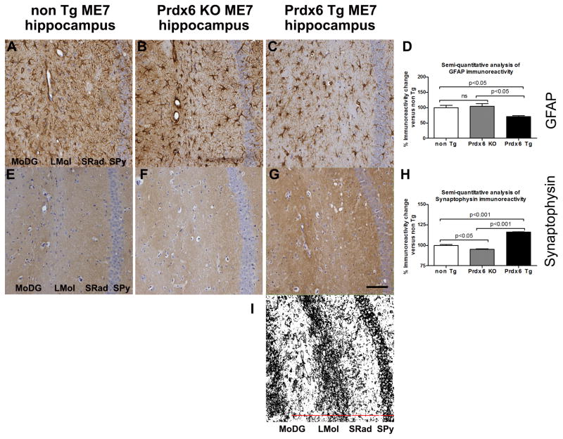
Representative images of astrogliosis and synaptopathy associated with increasing accumulation of misfolded PrPSc in nonTg ME7-, Prdx6 KO ME7- and Prdx6 Tg ME7-animals. (A–C) anti-GFAP antibody (shows increased numbers of activated astrocytes), (E–G) anti-Synaptophysin antibody shows loss of demarcated synaptic layers and increasing disorganisation of synaptic architecture and decreased expression of presynaptic marker protein in nonTg ME7- and Prdx6 KO ME7-animals while a more intact synaptic architecture is revealed in Prdx6 Tg ME7-animals. These markers of prion-related pathology were all exacerbated in Prdx6 KO ME7-animals compared to Prdx6 Tg ME7-animals. No neuropathological differences were seen in Prdx6 KO NBH- and Prdx6 Tg NBH-animals (not shown here). Minus antibody control staining was also carried out (not shown). (D) We used ImageJ software (http://rsb.info.nih.gov/ij) to semi-quantitatively score histopathology staining and express it as a percent IR in Prdx6 KO ME7 and Prdx6 Tg ME7 relative to IR in similarly stained sections from nonTg ME7-animals (n = 3). Using this approach there was a significant decrease in GFAP IR in the hippocampus of Prdx6 Tg ME7 compared to Prdx6 KO (p<0.001) and nonTg ME7-animals (p<0.001). (H) Synaptophysin IR was significantly reduced in the hippocampus of nonTg ME7 (p<0.001) and Prdx6 KO ME7 (p<0.001) compared to Prdx6 Tg ME7-animals, but there was also a difference in synaptophysin IR in nonTg ME7 compared to Prdx6 KO ME7-animals. 20x mag, and scale bar is 100 μm. (I) Serves to show the demarcation of the synaptic layers discussed in the text. DAB staining of the representative image in (G) was identified using ImageJ software, the Colour Deconvolution plugin was used to eliminate haematoxylin counterstain, and the image threshold was adjusted. Representative images are from coronal sections showing hippocampal region, stratum pyramidal (Spy), stratum radiatum (SRad), lacunosum moleculare of the CA1 (LMol), and molecular layer of the dentate gyrus (MoDG).

