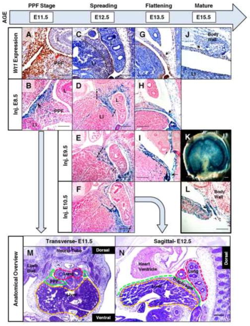
Fig. 1.
Wt1 contribution shifts in developing diaphragm mesothelium and mesenchyme in a time-dependent manner. Immunohistochemistry (A, C, G, J) was performed on tissue sections from wildtype mice at E11.5 to E15.5 to detect Wt1 protein localization in the developing diaphragm. Fate mapping was performed using whole-mount (B, D–F, H, K, L), or cryosectioned (I), X-gal stained Wt1CreERT2;R26Rlacz embryos harvested at E11.5 to E15.5 with recombination induced by tamoxifen injection at E8.5 (B, D, H), E9.5 (E, I), or E10.5 (F, K, L). Wt1 protein and Wt1-derived cells were localized to mesothelium of the PPF (arrows) in transverse sections at E11.5 (A, B). Injecting at different time points resulted in restricted patterns of lacz expression in sagittal sections at E12.5 (dotted lines) (D–F). Arrows indicate concentrated regions of Wt1 protein and Wt1 fate-mapped cells at E13.5 (G–I) and E15.5 (J–L). A whole X-gal stained diaphragm (K) at E15.5, injected at E10.5, is shown with a corresponding sagittal section (L). Low magnification H & E stained sections of representative wildtype embryos at E11.5 (M) and E12.5 (N) have relevant tissues and orientation labeled which is maintained throughout subsequent figures. Abbreviations: L, lung; Li, liver; PPF, pleuroperitoneal fold. The liver/diaphragm boundary is represented by a dotted line (C) where necessary. Scale bars: A, B, J, and L, 100 μm; C–I, M and N, 200 μm; K, 1 mm.

