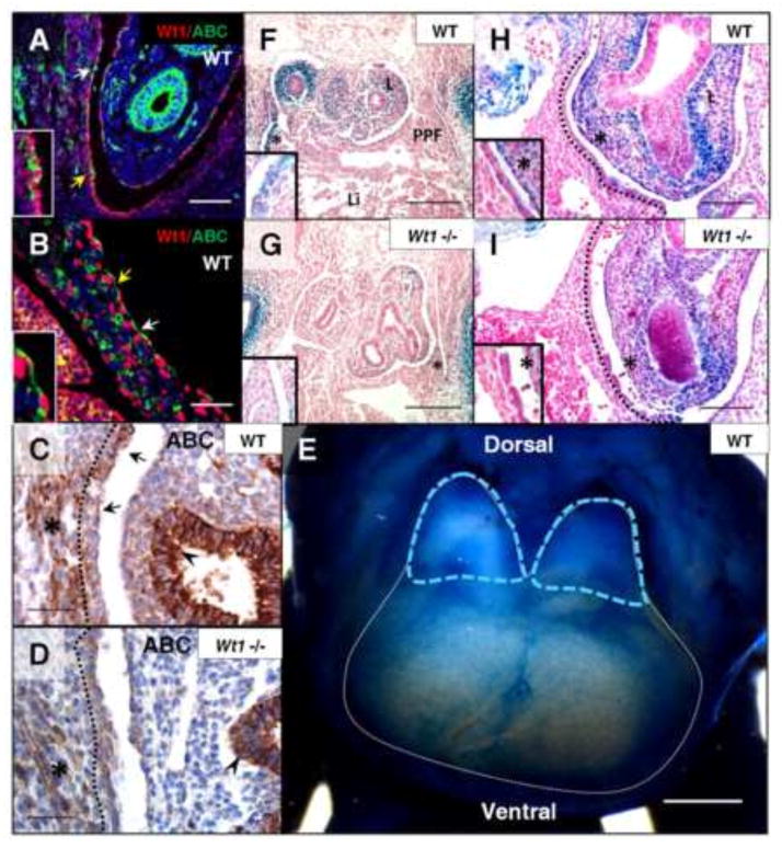
Fig. 2.
Loss of Wt1 results in reduced β-catenin expression in posterior diaphragm mesothelium. Wt1/Active β-catenin co-immunofluorescence was performed on sagittal sections of wildtype embryos at E12.5 (A) and E13.5 (B). Wt1 nuclear protein corresponds to regions of Active β-catenin staining in the mesothelial cell cytoplasm (arrows). Insets are cropped images of regions with yellow arrows. Active β-catenin immunohistochemistry (C, D) of E13.5 Wt1GFPCre wildtype and mutant (Wt1GFPCre/GFPCre) diaphragms labels regions of mesothelium (dotted line), indicated by arrows, as well as diaphragm muscle (asterisks) and lung epithelium (arrowheads). E12.5 whole mount X-gal stained Axin2lacz wildtype embryonic diaphragm (E) has been outlined in white, with blue dotted lines marking late PPF, dorsal and ventral sides are indicated. Axin2lacz;Wt1GFPCre embryos were harvested at E11.5 (F–I) and sectioned in transverse (F, G) and sagittal (H, I) orientation following whole mount X-gal staining. Insets are high magnification of region marked by asterisk. Abbreviations: ABC, Active β-catenin; L, lung; Li, liver; PPF, pleuroperitoneal fold. Scale bars: A, B, H and I, 100 μm; C and D, 50 μm; F and G, 200 μm; E, 0.5 mm.

