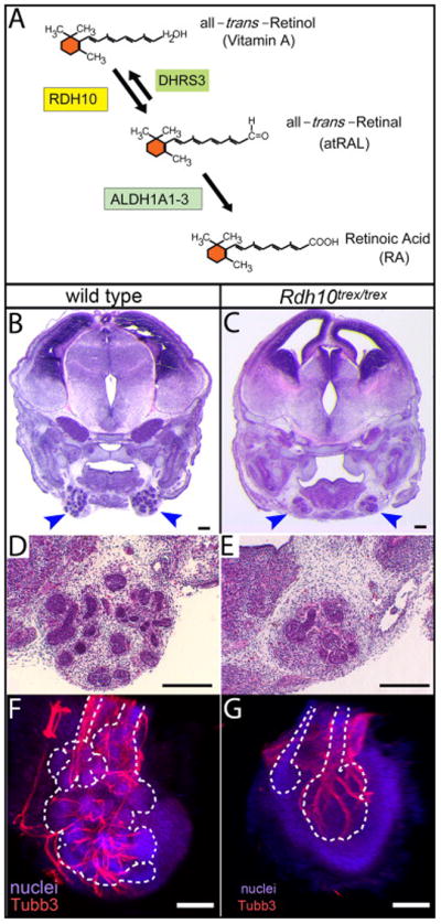
Figure 1. Deficient production of RA in Rdh10trex/trex embryos impairs development of the SMG.

(A) Vitamin A is converted into RA via two sequential enzymatic reactions. The first reaction, conversion of all-trans-Retinol (Vitamin A) to all-trans-Retinal (atRAL) is mediated within an embryo by RDH10. The first reaction is reversible, with the opposite reaction being mediated by DHRS3. The second reaction, the irreversible conversion of the intermediate atRAL to RA, is mediated by three aldehyde dehydrogenases, ALDH1A1, ALDH1A2, and ALDH1A3. Lack of RDH10 function results in severe RA deficiency and embryonic lethality usually prior to E11.5 (Sandell et al., 2007). (B–E) SMG development is impaired in Rdh10trex/trex embryos rescued to survive to E14.5 by maternal dietary supplementation with a minimal dose (40 μg) of the intermediate atRAL. Frontal paraffin sections through SMG of wild type and mutant embryos were stained with Hematoxylin and Eosin. SMG of 40 μg atRAL-rescued Rdh10trex/trex embryos (C, E) were substantially smaller than those of wild type littermates (B, D). Blue arrowheads indicate SMG. (F, G) Parasympathetic SMG ganglion and nerve development occurs in Rdh10trex/trex mutant embryos. Whole mount SMG from wild type (F) and Rdh10trex/trex mutant (G) embryos from 40 μg atRAL litters were immunostained for βIII neuronal tubulin (Tubb3), to reveal neurons of the parasympathetic ganglion and nerve, and with fluorescent nuclear stain Red Dot, to reveal all nuclei. For each gland a z-stack of confocal images was collapsed to form a single projection image. White dotted lines represent outline of gland epithelium visualized based on density of nuclear stain. Scale bars = 100 μm.
