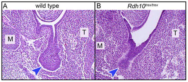
Figure 4. SMG initiation occurs in uncommon Rdh10−/− embryos surviving to E12.5 without atRAL supplementation.

In the absence of atRAL supplementation few Rdh10−/− embryos survive past E11.5. Rare example of surviving E12.5 Rdh10−/− mutant embryo has evidence of initiation of SMG morphogenesis. (A) SMG of wild type embryo at the E12.5 stage of development is an initial bud with open primary duct lumen connecting to the oral surface. Initial bulbous endbud is distinctly wider than primary duct. (B) SMG of mutant embryo appears as a thick dense epithelial invagination into the mesenchyme. Primary “duct” has no lumen, “endbud” is approximately same width as “duct”. M, Meckel’s cartilage; T, tongue; blue arrowheads indicate terminal endbud, or endbud-like region, of SMG epithelial invagination.
