Figure 7.

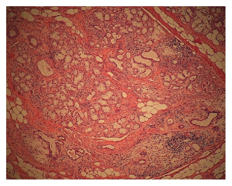
Histopathological examination (original magnification, ×40) reveals expansion of the acinus and duct, retention of mucus, atrophy of the acinus, and lymphocytic infiltration into the lesion of the bone cavity and sublingual gland.

Histopathological examination (original magnification, ×40) reveals expansion of the acinus and duct, retention of mucus, atrophy of the acinus, and lymphocytic infiltration into the lesion of the bone cavity and sublingual gland.