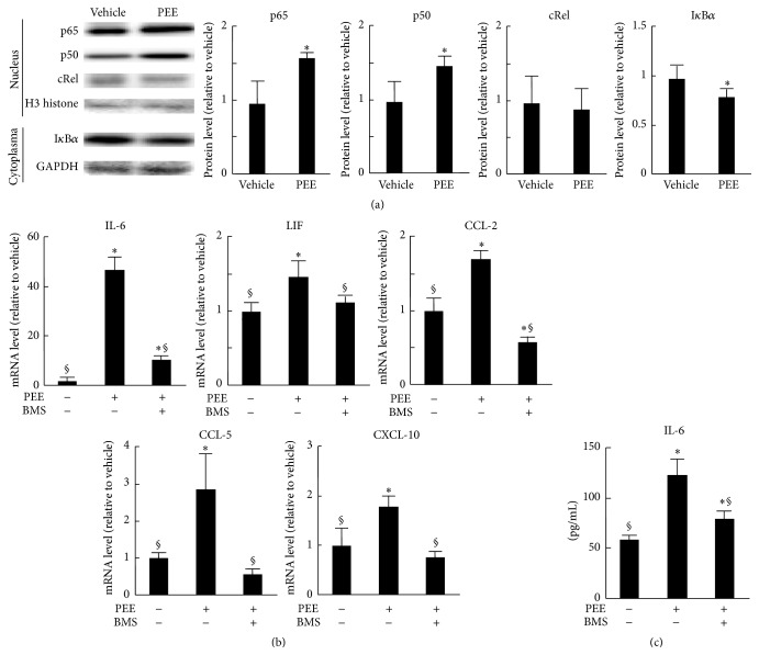
Figure 2.
Role of NF-κB p50 and p65 in propolis-elicited cytokine induction in C2C12 cells. (a) Western blotting analysis of nuclear p65, p50, and cRel and cytoplasmic IκBα from cells treated with 100 μg/mL PEE or vehicle for 3 h. H3 histone and GAPDH from nuclear and cytoplasmic protein, respectively, were used as loading controls. Left: representative blot images from three independent experiments. Right: quantitative data for band intensities. Data normalized to H3 histone (means ± SD of three samples). ∗ P < 0.05 versus vehicle-treated control cells. (b, c) Effects of 1 μM of an IKK inhibitor BMS-345541 on PEE-elicited cytokine induction. (b) Results of qRT-PCR analysis after treatment with PEE for 8 h. Data normalized to HPRT-1 mRNA (means ± SD of five samples). (c) Accumulation of IL-6 in culture medium 12 h after PEE application (means ± SD of eight samples). ∗ P < 0.05 and § P < 0.05 versus vehicle-treated control cells and PEE-treated, BMS-345541-untreated cells, respectively.

