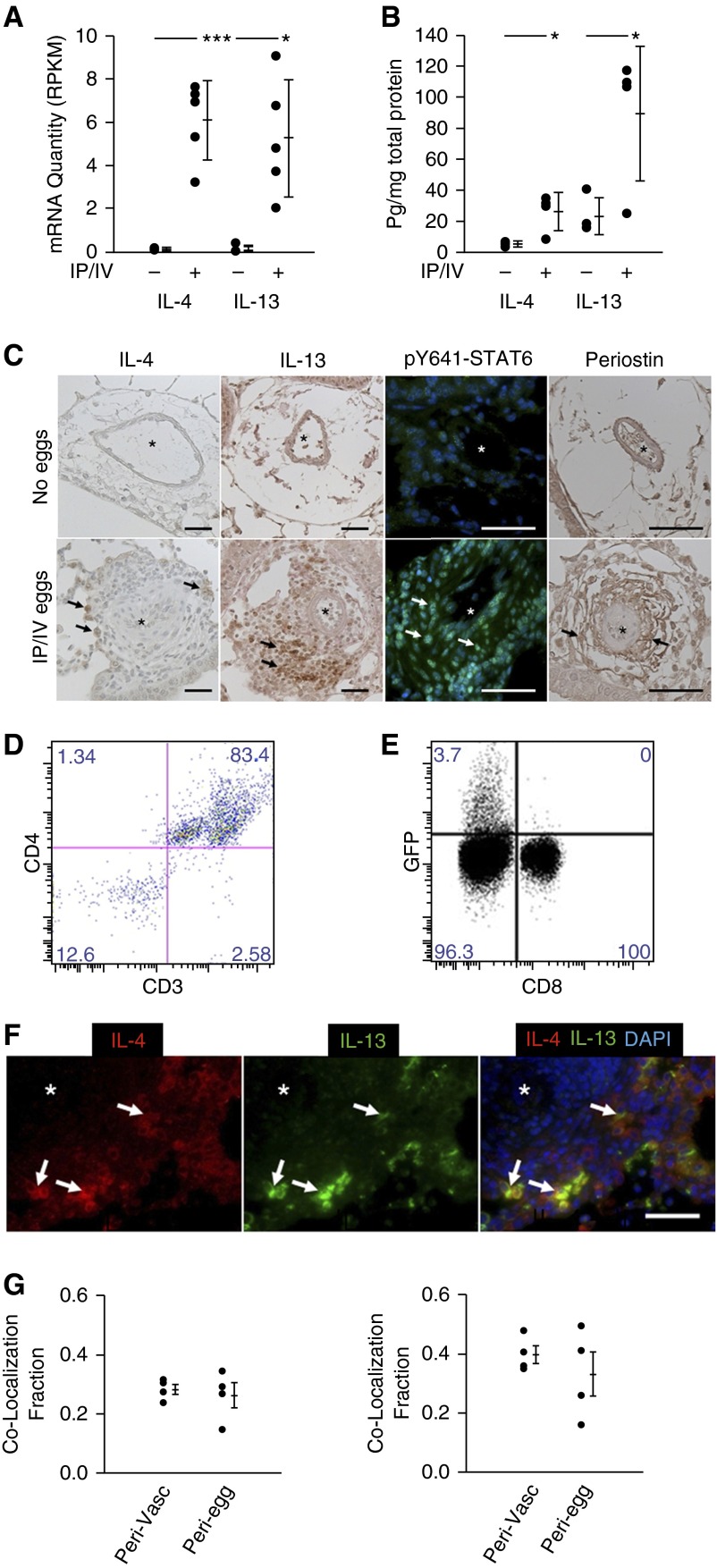
Figure 1.
Schistosoma-exposed mouse tissue demonstrates evidence of Th2 signaling. (A) Whole-lung quantity of IL-4 and IL-13 messenger RNA (mRNA) transcripts (n = 5 samples per group; RPKM; mean ± SD; t test, *P < 0.05; ***P < 0.005). (B) IL-4 and IL-13 whole-lung ELISA (mean ± SD; n = 4 samples per group; t test, *P < 0.05). (C) Immunostaining for IL-4, IL-13, phospho–signal transducer and activator of transcription factor 6 (STAT6) and periostin in Schistosoma-exposed and unexposed mouse lung tissue (asterisks, vessel lumens; arrows, representative positive stained cells; scale bars, 50 μm). (D) Flow cytometry analysis of GFP+ cells from intraperitoneal and intravenous (IP/IV) egg–exposed IL-4GFP mice for CD3+ and CD4+ cells. (E) Percentage of CD3+CD4+ and CD3+CD8+ cells in IP/IV egg–exposed mice positive for IL-4GFP (flow studies repeated at least three times). (F and G) Qualitative (F) and quantitative (G) colocalization of IL-4 and IL-13 using double immunoflorescence analysis (G, left, fraction of IL-4–positive area that colocalizes with IL-13; G, right, fraction of IL-13–positive area that colocalizes with IL-4; asterisk, vessel lumens; arrows, representative double-positive cells; scale bar, 50 μm; plots mean ± SD; n = 4 samples per group; the groups are perivascular/adventitia and periegg/granuloma images). DAPI = 4′,6-diamidino-2-phenylindole; GFP = green fluorescent protein; RPKM = reads per kilobase per million mapped reads.

