Figure 7.
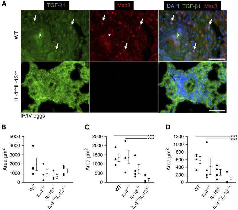
Macrophage density and transforming growth factor (TGF)-β1–macrophage colocalization is decreased in IL-4−/−IL-13−/− Schistosoma-exposed mice. (A) Representative images showing double immunofluorescence staining for TGF-β ligand and Mac3 (macrophage marker; images representative of n = 4/group; asterisks, vessel lumens; arrows, representative positive double-stained cells; scale bars, 50 μm). (B–D) Quantitative analysis of area in the adventitia that stains positive for TGF-β1 (mean ± SD; n = 4 specimens per group; analysis of variance [ANOVA], P = NS) (B), Mac3 (macrophages; mean ± SD; n = 4 specimens per group; ANOVA, P = 0.011; post hoc Tukey test, ***P < 0.005) (C), and colocalization of both TGF-β1 and macrophages (mean ± SD; n = 4 specimen per group; ANOVA, P = 0.011; post hoc Tukey test, ***P < 0.005) (D) in Schistosoma-exposed wild-type, IL-4−/−, IL-13−/−, and IL-4−/−IL-13−/− mice. DAPI = 4′,6-diamidino-2-phenylindole; IP/IV = intraperitoneal and intravenous; NS = not significant; WT = wild-type.

