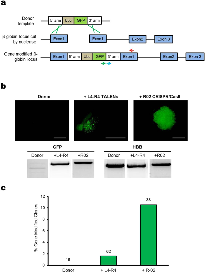
Figure 4. HDR-mediated gene modification in microinjected K562 cells.
(a) Diagram of GFP reporter system used to detect HDR. Nuclease cleavage and resection yields a substrate for HDR which may involve the use of exogenous β-Ubc-GFP donor, flanked by 5′ and 3′ homologous fragments of HBB sequence (top). The green lines indicate the HDR process with the donor template. Gene insertion was confirmed by PCR using primers for integrated GFP at the target locus, as shown by the green and red arrows (bottom). The HBB gene (control) was amplified using primers shown by the blue and red arrows, which bound downstream the 3′ homologous region in HBB. (b) Fluorescence microscopy images of clones derived from single cells microinjected with β-Ubc-GFP donor with or without L4-R4 TALENs or R02 CRISPR/Cas9. Bottom of images are PCR results of integrated GFP or HBB (control) for clones. Scale bar corresponds to 500 μm. (c) The percentage of clones with HDR-mediated gene modification confirmed by both PCR and fluorescence microscopy. The number of single cell clones analyzed is shown above each bar.

