
Figure 5. Gene insertion in nucleofected cells.
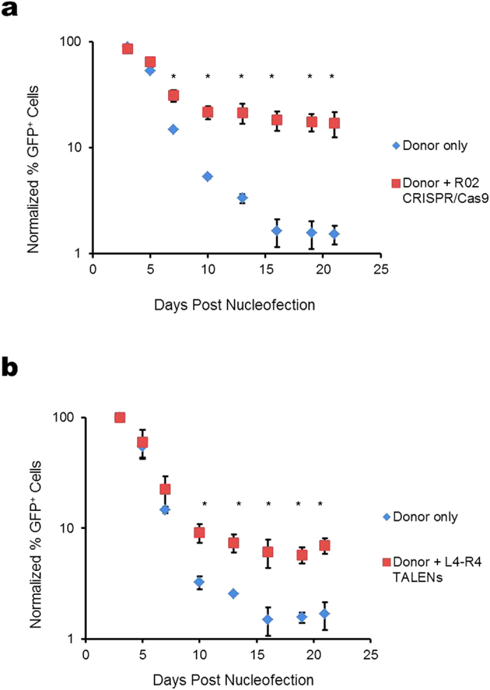
The percentage of GFP-positive cells was quantified using flow cytometry during a 21-day culture after nucleofection with β-Ubc-GFP donor with and without (a) R02 CRISPR/Cas9 or (b) L4-R4 TALENs. The plots show normalized percentage of GFP-positive cells at specified days post nucleofection. Asterisks indicate significant difference between donor only and donor plus nuclease at specified days.