
Figure 8.
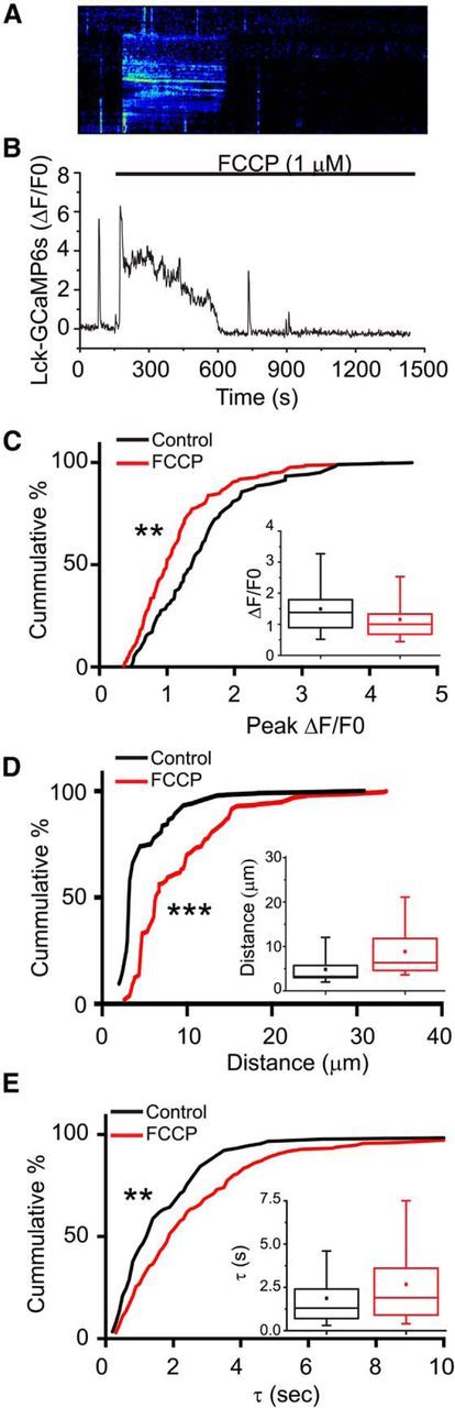
Mitochondria regulate Ca2+ signaling within astrocyte processes. Astrocytes in organotypic cultures of rat hippocampus were transfected with a membrane-targeted-mCherry fluorescent protein driven by a GFAP promoter (GFAABC1D-mCherry) and the genetic Ca2+ indicator LCK-GCaMP5G. Slices were continuously superfused with oxygenated-aCSF. FCCP (1 μm) was applied by superfusion as indicated by application bar. Representative kymograph (A) and fluorescence trace (B) from an astrocyte processes treated with the proton ionophore (FCCP; 1 μm). The amplitudes (C, ΔF/F0), distance traveled (D, μm), and kinetics (E, tau, seconds) of the individual Ca2+ transients were quantified before (black) and after application of FCCP (red), and the results are depicted as both cumulative probability distributions and box-and-whisker plots (inset). Error bars indicate 5%–95% range. Shoulders of boxes indicate 25%–75% intervals. Median of data is highlighted by horizontal line. **p < 0.001 (Kolmorogov–Smirnov test). ***p < 0.0001 (Kolmorogov–Smirnov test). n = 90 and n = 208 spikes (81 processes, 5 cells) for control and FCCP-treated conditions.