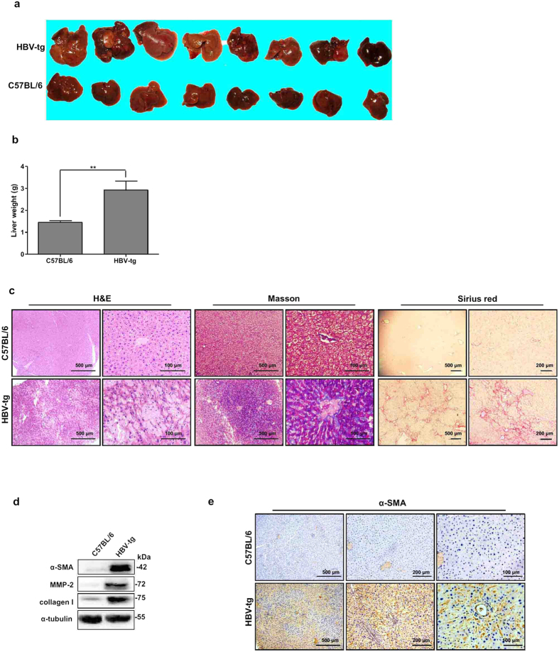
Figure 6. Spontaneous liver fibrosis in HBV-tg mice.
(a) Morphology of liver and (b) liver weight of 12-month-old C57BL/6 and HBV-tg mice (n = 8). **P < 0.01. (c) H&E, Masson′s trichrome, and Sirius red stainings of liver tissues from 6-month-old C57BL/6 and HBV-tg mice. (d) Western blot analysis of α-SMA, MMP-2, and collagen I expressions in liver tissues from 6-month-old C57BL/6 and HBV-tg mice. (e) Imm-unohistochemistry detected α-SMA expression in liver tissues from 6-month-old C57BL/6 and HBV-tg mice.

