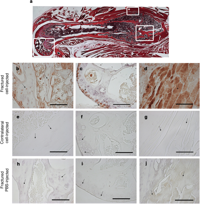
Figure 5. RFPpos CH cells actively participate in tissue healing.
(a) Serial hystology sections (magnification 5X) from the resulting fracture callus formed 24 days after the osteotomy. #hard callus; °muscle tissue surrounding the callus; *knee region. (b,e,h) Representative anti-RFP immunostaining of the hard callus region derived from fractured and cell-injected mouse (b), corresponding region of the contralateral paw of the same mouse (e), and the hard callus region derived from the fractured and PBS-injected mouse (h). Black arrows indicate osteoblasts within the bone matrix. (c,f,i) Representative anti-RFP immunostaining of the knee region derived from fractured and cell-injected mouse (c), contralateral paw of the same mouse (f), and fractured and PBS-injected mouse (i). Black arrows indicate articular chondrocytes. (d,g,j) Representative anti-RFP immunostaining of the muscle tissue derived from fractured and cell-injected mouse (d), contralateral paw of the same mouse (g), and fractured and PBS-injected mouse (j). Black arrows indicate muscle fibers. Magnification 40X, scale bar 100 μm.

