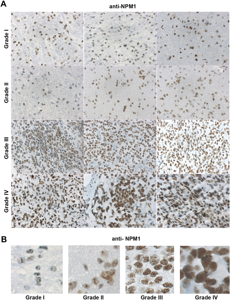
Figure 1. Detection of NPM1 in astrocytic gliomas.
(A) Immunohistochemical staining of NPM1 (brown) in astrocytic glioma tumors of grades I, II, III and IV. Three tumor samples from different patients are shown for each grade. Obj. 20x. (B) Representative zoom-in micrographs showing NPM1 staining in tumor samples of different grades (I-IV).

