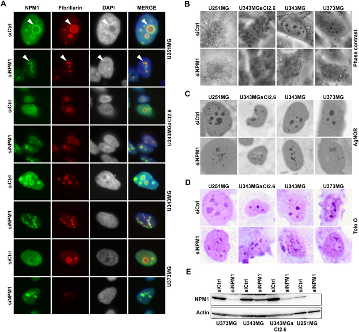
Figure 3. Knockdown of NPM1 in glioma cells alters nucleolar morphology.
(A) IF staining of NPM1 (green) and fibrillarin (red) in glioma cell lines U343MGa Cl2:6, U373MG, U343MG, and U251MG treated with siNPM1 for 6 days. Nuclei were counterstained with DAPI. Arrowheads point at nucleoli. (B) Phase contrast micrographs of control and of cells depleted of NPM1. (C) AgNOR staining of control and NPM1 depleted glioma cells. (D) Acidic Toluidine Blue O staining of glioma cells. Nucleoli appear as darker stained than the surrounding nucleoplasm. (E) IB analysis of NPM1 levels in different glioma cell lines as indicated after siNPM1 treatment for 6 days. β-actin served as loading control.

