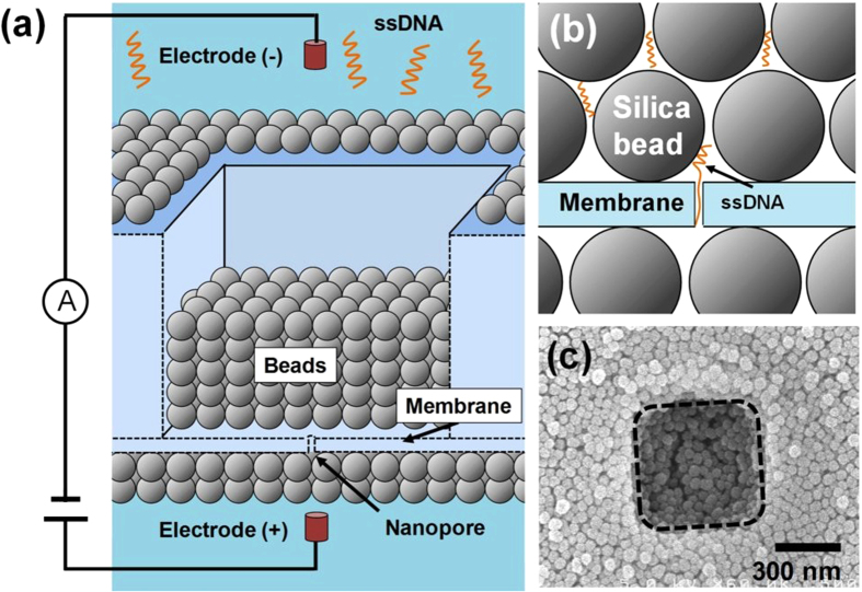
Figure 1. Nanopore with nanometre-sized bead nanostructure.
(a) Schematic of solid-state nanopore with nanometre-sized bead structure coated on the membrane. (b) Schematic showing interaction between ssDNA and the silica bead around a nanopore. (c) Typical top-view scanning electron microscope image of the bead-coated substrate. The dashed line shows the square area that is the thinnest part of the membrane. (The thickness of the membrane: 20 nm, the diameter of the beads: 50 nm, the width of the square area: 500 nm, the base length of ssDNA: 60-mer.).

