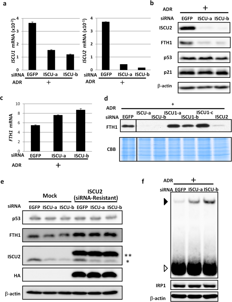
Figure 4. Regulation of FTH1 expression by ISCU.
(a) Twenty-four hours after transfection of each siRNA, HCT116 cells were treated with ADR. After 36 h, cells were collected, and qPCR analysis was performed. siRNA against EGFP was used as a control. ACTB was used to normalize the expression levels. Error bars represent the S.D. (n = 3). (b) Twenty-four hours after transfection of each siRNA, HCT116 cells were treated with ADR. After 36 h, cell extracts were subjected to western blot analysis. siRNA against EGFP was used as a control. β-actin is shown as a loading control. (c) Twenty-four hours after transfection of each siRNA, HCT116 cells were treated with ADR. After 36 h, cells were collected, and qPCR analysis was performed. siRNA against EGFP was used as a control. ACTB was used to normalize the expression levels. Error bars represent the S.D. (n = 3). (d) Twenty-four hours after transfection of each siRNA, HCT116 cells were treated with 2 μg/ml of ADR. After 36 h, cells were harvested for western blot assay. CBB staining is shown as a loading control. ISCU1-a, ISCU1-b, ISCU1-c, or ISCU2 indicate siRNA specific for ISCU1 or ISCU2, respectively. (e) Twenty-four hours after transfection of the mock or ISCU2 expression vector, HCT116 cells were treated with siRNA. After 24 h, HCT116 cells were incubated with 2 μg/ml of ADR. Cells were collected for western blot assay 36 h after ADR treatment. β-actin is shown as a loading control. **Exogenous ISCU2 (HA-tagged ISCU2). *Endogenous ISCU2. (f) RNA-EMSA using a biotin-labelled probe containing the IRE in the 5′ UTR of FTH1 mRNA. Twenty-four hours after transfection of each siRNA, HCT116 cells were treated with ADR. After 36 h, cytosolic cellular fractions were incubated with probe for 30 min, and electrophoresis mobility shift assays were performed (upper). The open arrowhead indicates free probe, and the closed arrowhead indicates the protein-RNA complex. Cytosolic fractions were also subjected to western blot analysis using an anti-IRP1 or anti-β-actin antibody (lower).

