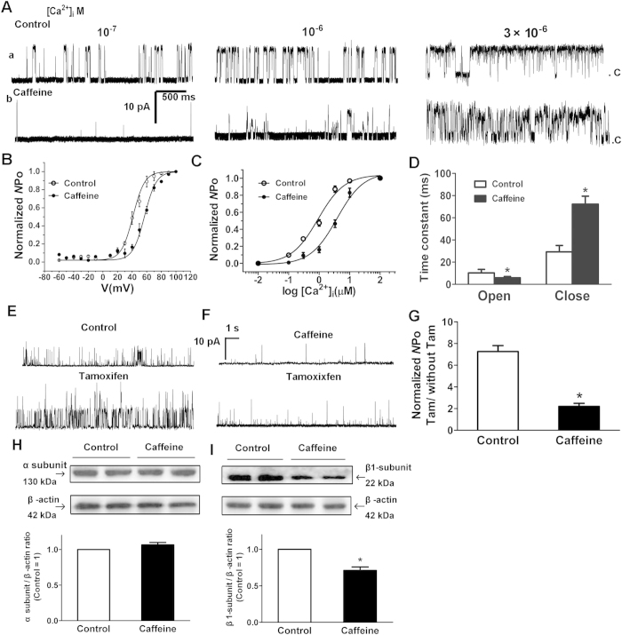
Figure 5. The effects of prenatal caffeine on biophysical properties of BKCa channels on myocytes from mesenteric arteries (MA).
(A) Representative single BKCa channel records in inside-out patches (HP = + 40 mV) from the control (a) and caffeine (b) myocytes exposed to increasing [Ca2+]i. (B) Voltage dependence of BKCa channels in [Ca2+]i at 10−6 mol/L. The line was drawn according to the best fits with the Boltzmann equation. (C) The effect of prenatal caffeine on Ca2+ sensitivity of BKCa channels. The data points were fitted with the Hill equation to obtain the calcium concentration necessary to open half of the channels (Kd) and the Hill coefficient (ηH). (D) Summary of time constant for open and close state of BKCa channels ([Ca2+]i = 10−6 mol/L, HP = + 40 mV; n = 30 cells, 7 animals/each group). (E–F) Representative single BKCa channel recording in inside-out patches (HP = + 40 mV, [Ca2+]i = 10−7 mol/L) from the control (E) and caffeine (F) myocytes exposed to 10−6 mol/L tamoxifen (Tam). (G) Bar plot summarizes the mean ± SEM fold change in the Normalized NPo of BKCa channels after the application of Tam. (n = 30 cells, 7 animals/each group). (H–I) Protein expression of BKCa α (H) and β1-subunit (I). The α and β1-subunit expression in the caffeine MA was presented relative to control vessels (n = 6 each group). *P < 0.05, control vs. Caffeine. Gels were treated under the same experimental conditions with the control and experimental groups treated together.

