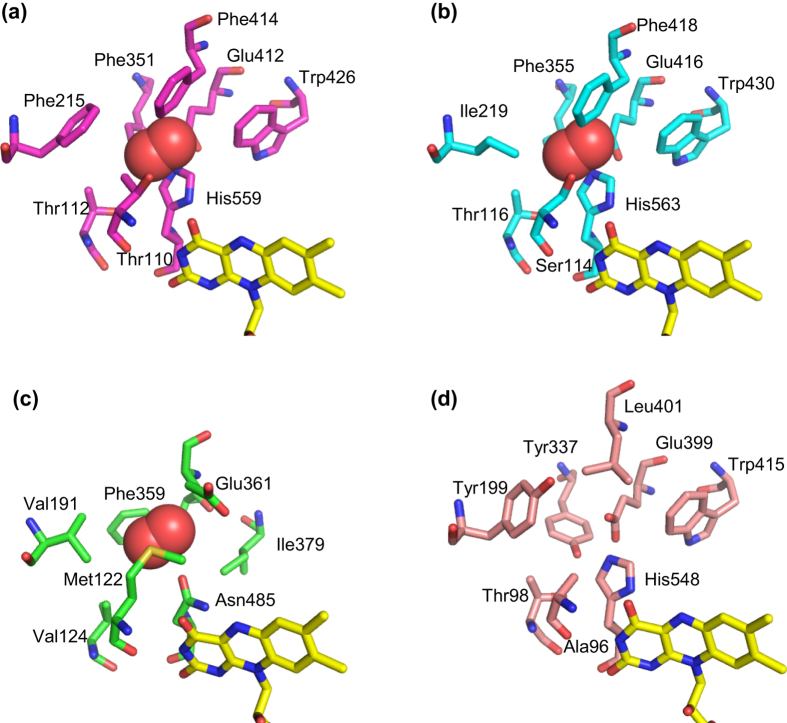
Figure 6. Comparison of residues responsible for the oxygen binding of AnGOx (1CF3), PaGOx (1GPE), ChOx (1MXT), and corresponding residues in AfGDH.
(a) AnGOx (protein, magenta; FAD, yellow). (b) PaGOx (protein, cyan; FAD, yellow). (c) ChOx (protein, green; FAD, yellow). (d) AfGDH (protein, pink; FAD, yellow). Oxygen molecules are represented as red spheres.

