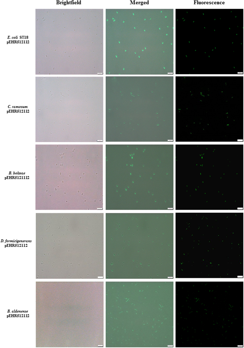
Figure 4. Analysis of transconjugants carrying pEHR512112 by fluorescence microscopy.
The transconjugants were recovered and purified on M2SC based medium. Colonies were re-suspended in sterile anaerobic diluent and individual cells were visualised using an Olympus BX 63 microscope. Images were captured using the Olympus cellSens modular imaging software platform and processed using the ImageJ software package. A scale bar of 10 μm is included for reference.

