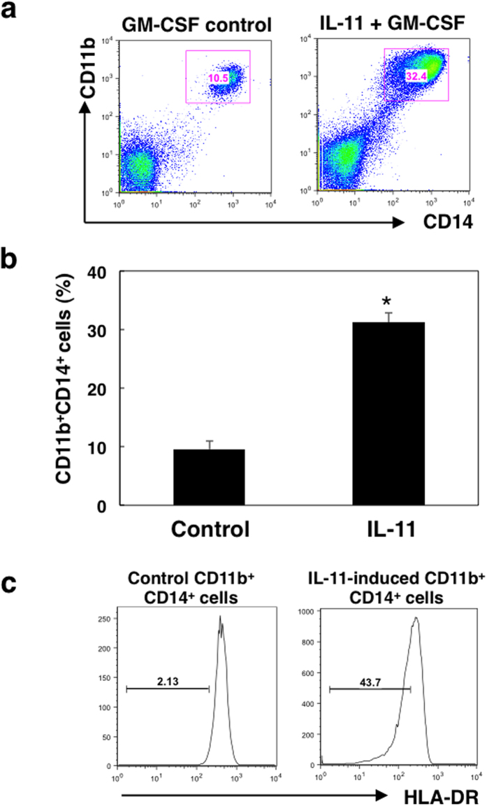
Figure 1. Effect of IL-11 on differentiation of PBMCs into CD11c+CD11b+ monocytic cells. PBMCs collected from blood of healthy donors were cultured with IL-11 (10 ng/ml) and GM-CSF (50 ng/ml) or GM-CSF alone for 7 days, and surface markers were analysed by flow cytometry.
(a) Representative dot plots of CD11b+ and CD14+ cells. (b) Means and SDs for the data from three independent experiments are shown. *p < 0.05, compared with control, two-sided Student’s t test. (c) Surface expression levels of HLA-DR on CD11b+CD14+ cells were evaluated by flow cytometry. Bars represent HLA-DR negative and/or low populations. The representative of three independent experiments is shown.

