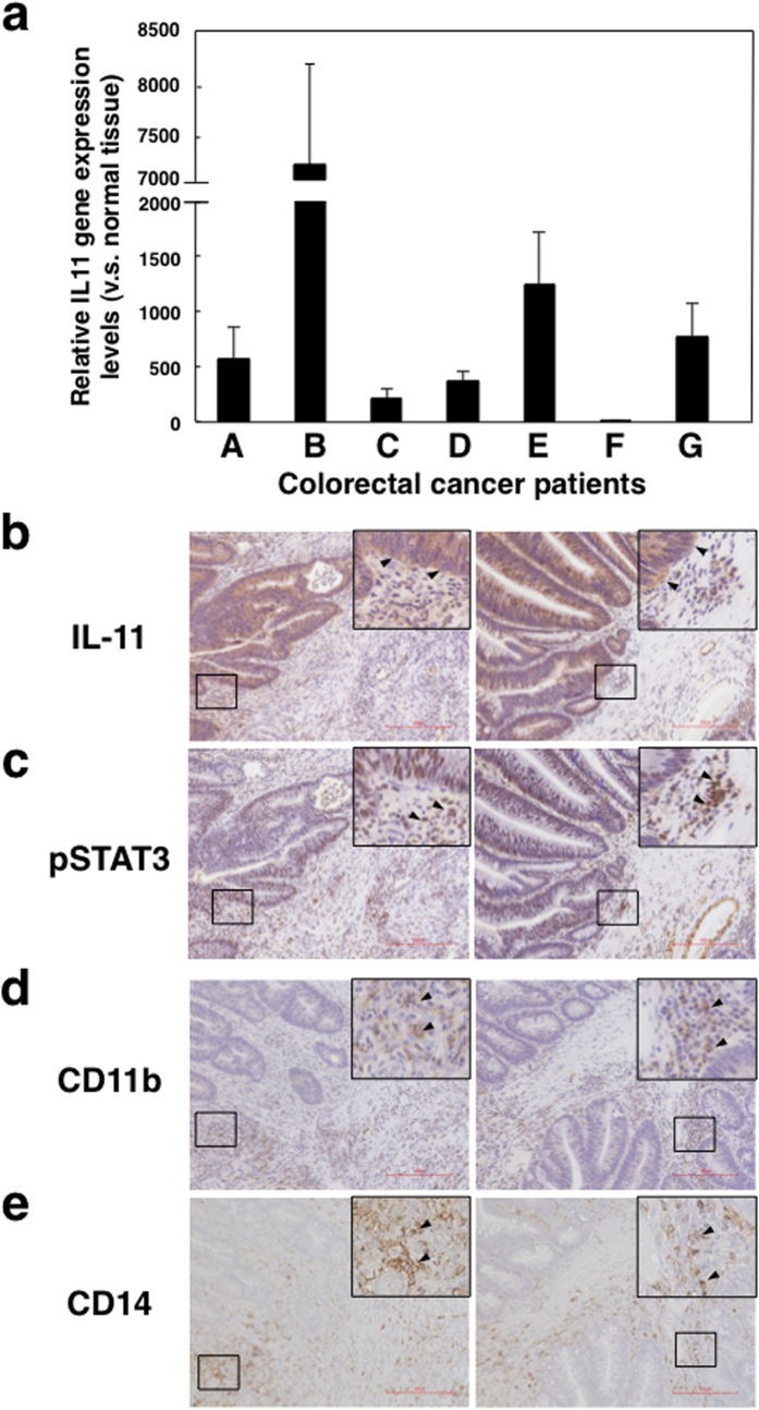
Figure 5. IL-11 and phosphorylated STAT3 expression in tumour microenvironments of colorectal cancer patients.
(a) Normal and tumour tissues were collected from the specimens of seven colorectal cancer patients. Gene expression levels of IL-11 and GAPDH in normal and tumour tissues were determined by quantitative PCR. IL-11 gene expression in each sample was normalized to levels of GAPDH. Relative IL-11 gene expression levels of tumour tissues against normal tissues were calculated. The means and SDs of the data from three independent experiments are shown. *p < 0.05, compared with control, two-sided Student’s t test. IL-11 (b), pSTAT3 (c), CD11b (d), and CD14 (e) protein expressions in tumour tissues of colorectal cancer patients were detected by immunohistochemistry. Scale bar is 200 μm for all panels. Representative photos including magnified views and allows of patient B and E are shown.

