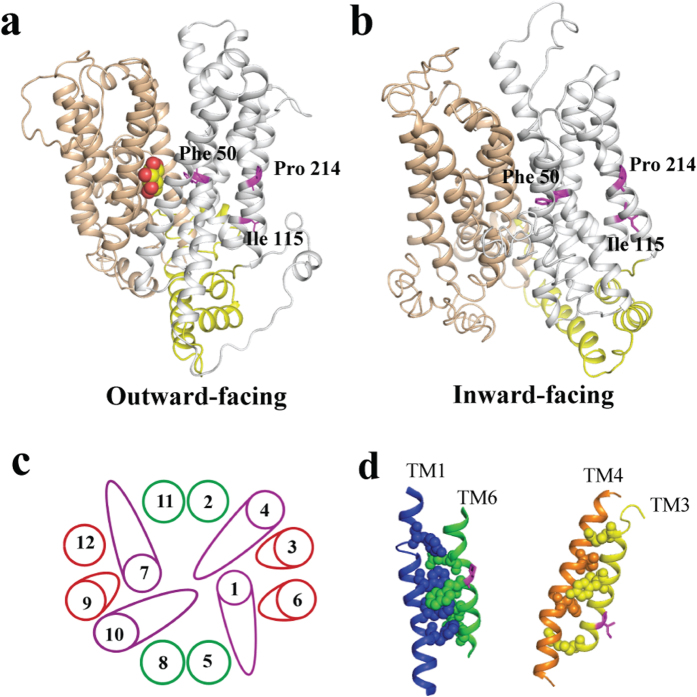
Figure 3. Modelled structures of Stp1 and its putative helix-packing arrangement.
Modelled structures were performed with the crystal structures of XylE bound to D-glucose (a) and GLUT1 in an inward-open state (b) as the template, respectively. The N and C domains are coloured grey and tan, respectively, and intracellular helices connecting these two domains are coloured yellow. D-glucose is indicated by the spheres. (c) Putative helix-packing arrangement viewed from the extracellular surface. (d) Putative stacking of TMs 6 and 1 (left) and TMs 3 and 4 (right). Residues with putative interactions are shown as spheres. Phe 50, Ile 115 and Pro 214 are represented in magenta.

