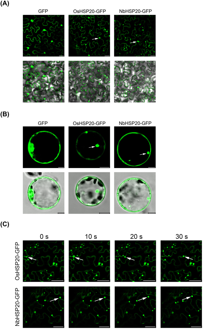
Figure 3. Sub-cellular localization of OsHSP20 and NbHSP20 proteins.
(A) GFP fluorescence in N. benthamiana leaf epidermal cells agroinfiltrated with pCV-GFP-N1, pCV-OsHSP20-GFP and pCV-NbHSP20-GFP, respectively. The results were observed 48 h after infiltration. Scale bar, 25 μm. (B) GFP fluorescence in rice protoplasts transfected with pCV-GFP-N1, pCV-OsHSP20-GFP and pCV-NbHSP20-GFP, respectively. The results were observed 18 h after transfection. Scale bar, 5 μm. The white arrow points to a granule. The fluorescence and merged images are depicted in the upper and lower panels, respectively. (C) Images recording the movement of OsHSP20-GFP or NbHSP20-GFP in N. benthamiana epidermal cells. In each local field (upper and lower), four sequential pictures detecting green fluorescence were taken at 0, 10, 20 and 30 s. The mobile GFP granules are marked with white arrows. Scale bar, 50 μm.

