Figure 2.
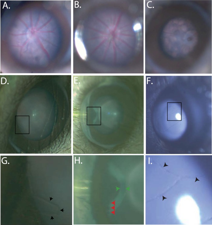
Bright field in vivo images demonstrating the RIS depth of field and imaging capabilities. Representative fundus images of a C57BL6(N) (6 months) (A), A wild-type human RHO on the mouse RHO knockout background (2HRho//1T/1T; ∼4 months of age) (B); Mouse RHO knockout (129R-; ∼4 months of age). (C) Regions of interest, demonstrating depth of field of the RIS, are boxed in images (D–F) are expanded in images (G–I) respectively, and show fine anatomical structures which can be identified. Images were enhanced for contrast and brightness to improve the fine details in images (A, B, G, I). Images from an A1 mouse (∼1 month of age) of the anterior chamber, both the cornea and iris are clearly visible, even the smallest of blood vessels are visible protruding from the edge of the iris (black arrow heads) (D, G). The pars plana can be imaged with scleral depression allowing visualization of ciliary body processes (red arrow heads) and the retinal thickness can be visualized highlighted by the (green arrow heads) (E, H). Visualization of the hyaloid vessels (black arrow heads) is shown in (F, I) in a C57BL6(N); ∼2-week old). Imaging of the mouse retina including optic nerve head and vasculature and retinal pigmentation can be clearly visualized (A–C). These images demonstrate the depth of view of the internal contents of the mouse eye provided by the RIS and the resolution capabilities of this system. The field of view allows the entire cornea to be imaged, but is more limited when imaging the retina due to the small diameter of the dilated mouse pupil, the effects of the mouse lenticular optics, and the long working distance of the instrument. Imaging of the entire retinal surface can be accomplished by manipulation of the eye or animal.

