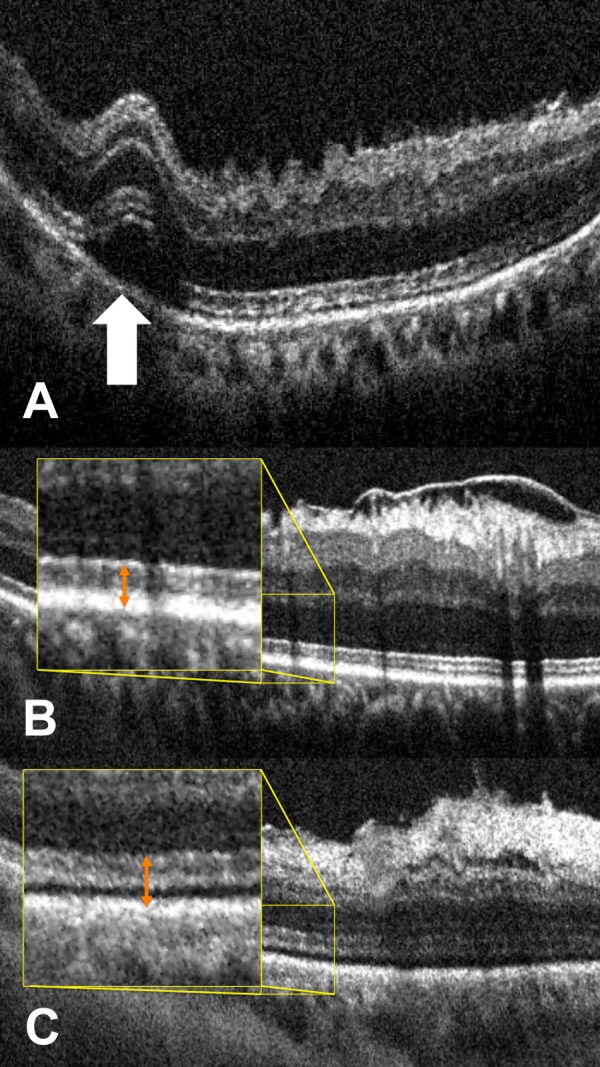
Figure 1.

Macroarchitectural and microarchitectural alterations on iOCT following membrane peeling. (A) Postpeel iOCT B-scan revealing full-thickness retinal elevation at area of engagement demonstrating macroarchitectural change (white arrow). (B) Prepeel iOCT B-scan showing ERM and typical EZ-RPE height (inset, orange double arrow). (C) Postpeel iOCT demonstrating removal of ERM with associated microarchitectural alterations with expansion of the EZ-RPE (inset, double orange arrow).
