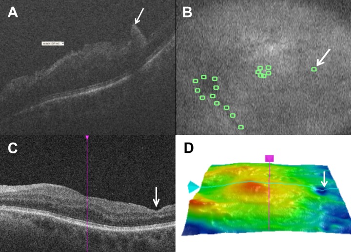
Figure 3.
Longitudinal assessment of retinal anatomy associated with macroarchitectural alterations. (A) Intraoperative optical coherence tomography following membrane peeling with macroarchitectural alteration identified as a possible full-thickness retinal elevation (white arrow). (B) Summed voxel projection with manual landmark identification and corresponding architectural alteration identification (white arrow). (C) Postoperative optical coherence tomography with inner retinal thinning in the area corresponding to previous retinal elevation. (D) Postoperative tomographic retinal thickness map with focal area of thinning (white arrow) in the temporal retina corresponding to area of intraoperative macroarchitectural changes.

