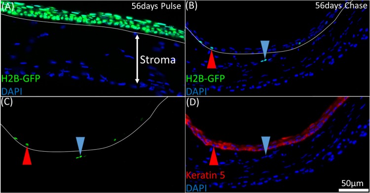
Figure 2.
LRCs in the H2B-GFP/K5tTA mouse were localized to the limbus epithelium and infrequently, the anterior limbal stroma. (A) At 56 days pulse, all keratin 5+ corneal epithelial cells express nuclear GFP tagged to histone H2B. (B, C) A 2 μm BMMA cross-section of the H2B-GFP/K5tTA mouse limbus after 56 days doxycycline chase. GFP+ cells were primarily localized to the limbus epithelium (red arrow); however, on rare occasion, they were observed in the limbus stroma with varying fluorescence intensities (blue arrow). (D) Keratin 5 staining (B, C) shows that the GFP+ stromal cells are Keratin 5−. This suggests that migration of limbal epithelial cells may occur or there is ectopic expression of keratin 5.

