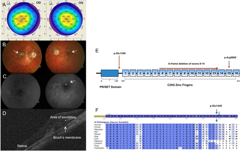
Fig. 2.

A case of PRDM5-associated disease presenting with myopic choroidal neovascularization. a. Corneal topography of right eye (OD) and left eye (OS) in patient P4 demonstrating marked corneal thinning, with central corneal thickness measuring 276 μm right eye and 281 μm left eye. b. Retinal photograph of RE (upper left panel) and LE (upper right panel) demonstrating evidence of disciform-like scarring in the macular region of the RE (arrow), and active choroidal neovascularization in the LE (arrow) leading to retinal exudation. Disc pallor and choroidal thinning are also present, related to the high refractive error of P4. c. Two fluorescein angiogram images of the LE taken at the time of presentation. Early phase (left panel) and late phase (right panel). An area of exudation indicative of vascular leakage into the retina is evident, suggestive of predominantly classic choroidal neovascularization. d. Optical coherence tomography scan of the LE at baseline demonstrating the presence of vascular exudation into the retina (*). e. Schematic of PRDM5 protein showing the novel mutation p.Glu134*; PRDM5 deletion exons 9–14; and p.Arg590*. f. Amino acid conservation in 11 species upstream and flanking the mutation p.Glu134*. The alignment was generated using Alamut v2.3 (Interactive Biosoftware, 2013)
