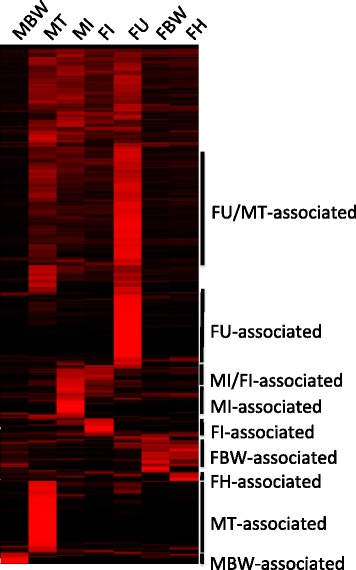
Fig. 2.

Comparison of D. immitis gene expression profiles between D. immitis tissues. Data from biological replicates were combined prior to clustering. The color scale ranges from black (no expression) to red (very high expression). Since Additional file 2: Dataset S1 lists all currently annotated D. immitis protein coding genes, only genes expressed in at least one tissue are shown (n = 11,640). Each gene is represented by a single row of boxes. Tissue-specific clusters used for tissue-specific functional analysis are indicated (black bars)
