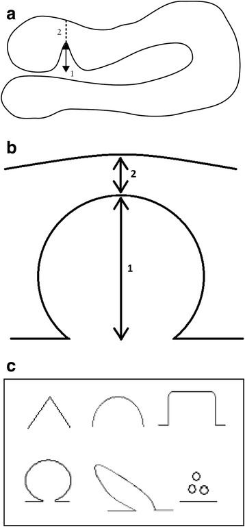
Fig. 3.

Niche measurement during sonohysterography in the sagittal plane (a), transversal plane (b) with the thinnest residual myometrium and niche shape will be registered in both planes (c). a Measuring a niche in the sagittal plane. Schematic drawing demonstrating how to measure a niche in the sagittal plane. The depth of the niche is measured from the usual limit of the uterine cavity until the apex of the niche (1), the residual myometrium from the apex of the niche until the serosa (2). a is an adapted figure of the one that was published by Bij de Vaate et al. 2011 [5]. b Measuring a niche in the transversal plane. Schematic drawing demonstrating how to measure a niche in the transversal plane. The depth of the niche is measured from the usual limit of the uterine cavity until the apex of the niche (1) and the residual myometrium from the apex of the niche until the serosa (2). c Niche shape. Schematic diagram demonstrating classification used to assess niche shape as published by Bij de Vaate et al. 2011: triangle, semicircle, rectangle, circle, droplet and inclusion cysts [5]
