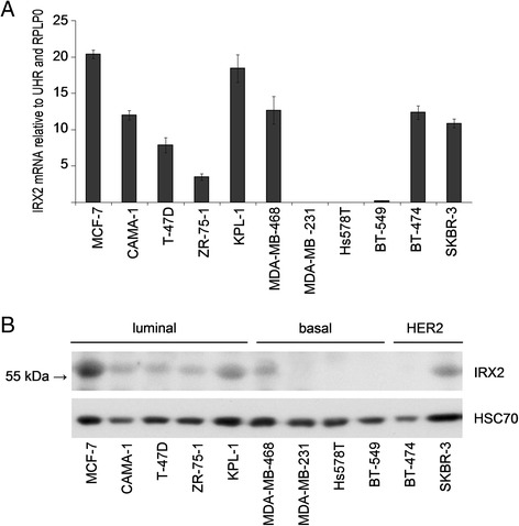
Fig. 1.

Analysis of IRX2 mRNA and protein expression in breast cancer cell lines. a Quantitative gene expression analysis of IRX2 in different breast cancer cell lines. The data shown are the average fold change normalized to RPLP0 and UHR expression of three independent experiments; the error bars represent the standard deviation of the mean. b IRX2 protein expression in the same panel of human breast carcinoma cell lines was determined by Western blot analysis using a polyclonal RAI2-specific antibody that recognizes an internal IRX2 epitope. Equal loading was demonstrated using an antibody recognizing HSC70
