Fig. 7.

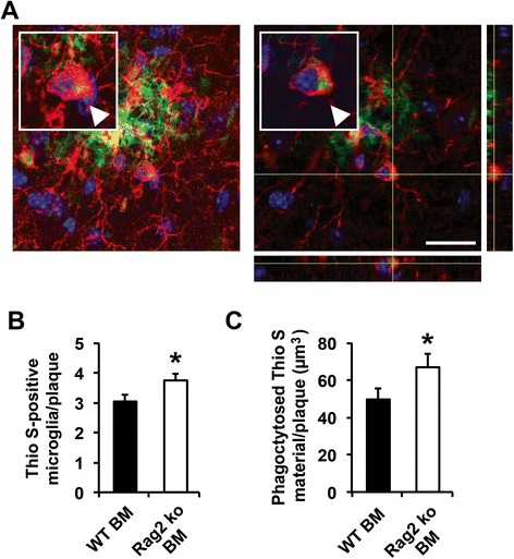
Increased number of Aβ-phagocytosing microglia in the Rag2 ko BM-reconstituted PSAPP mice. a Representative maximum projection confocal image (left panel) of a plaque-associated microglial cell containing Thioflavin S-positive Aβ material (arrowhead). Image shows microglia (Iba1, red) and amyloid deposits (Thioflavin S, green). Nuclei were counterstained by DAPI (blue). Orthogonal views of z-stack images are shown in the right panel. Scale bar = 20 μm (b) significantly increased number of amyloid plaque-associated Iba1-positive microglial cells containing Thioflavin S-positive Aβ material in the Rag2 ko BM-recipient PSAPP mice. *p < 0.05. n = 9 mice per group (on average 12 plaques per mouse analyzed) (c) significantly increased total volume of phagocytosed Aβ material in the Rag2 ko BM-reconstituted PSAPP mice in comparison to the WT BM-recipient control mice. *p < 0.05. n = 9 mice per group (on average 12 plaques per mouse analyzed)
