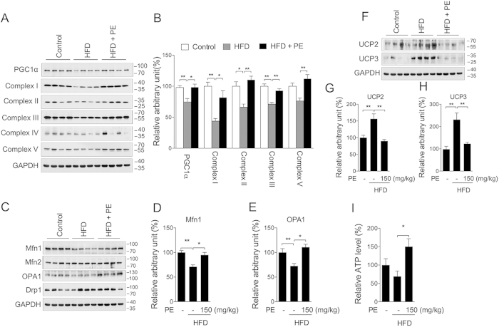
Figure 6. PE promotes mitochondrial remodeling in hearts of rats fed a HFD.
Extracts were prepared from the rat heart to assess total protein content. The mitochondrial content was assessed by measuring PGC-1α and the mitochondrial complexes (A) western blot image; (B) arbitrary units analysis). The mitochondrial dynamic status was assessed by measuring the Mfn1, Mfn2, OPA1 and Drp1 protein expression levels (C) western blot image; arbitrary units analysis of (D) Mfn1 and (E) OPA1). The UCP2 and UCP3 protein levels (F) western blot image; arbitrary units analysis of (G) UCP2 and (H) UCP3) as well as the tissue ATP content (I) were also analyzed. The values are presented as the means ± S.E.M. (n = 10); *p < 0.05, **p < 0.01.

