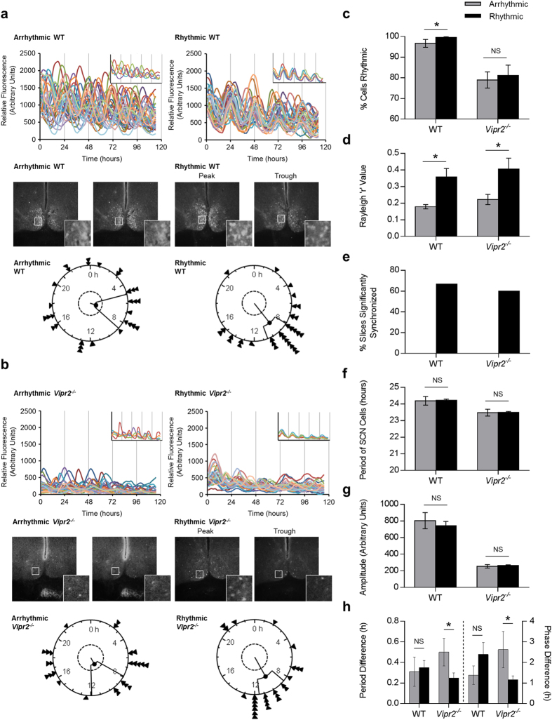
Figure 3. Behavioral rhythmicity in WT and Vipr2−/− mice is associated with increased intercellular synchrony in the SCN. a-b (upper panels).
Composite rhythm plots show per1::eGFP profiles for 30 individual SCN cells in two brain slices from each genotype. Insets show a reduced selection of cells to aid visualization of synchrony and highlight differences in synchrony between cells within slices. (a–b) (middle panels): Photomicrographs showing peak and trough expression of per1::eGFP for rhythmic slices and representative images from equivalent timepoints for arrhythmic slices. Insets show the regions indicated by white boxes at higher magnification (note synchronized peak and trough expression for SCNs from rhythmic mice and lack of coherent day-night differences for SCNs from arrhythmic mice). (a,b) (lower panels): Rayleigh plots showing time of peak per1::eGFP expression of rhythmic SCN cells analyzed within single slice cultures. Arrow heads indicate the time of peak fluorescence for individual cells, the length of the central line indicates the degree of synchrony between the times of peak expression of individual cells (quantified as Rayleigh R; longer line indicates greater synchrony) and the inner broken ring shows the threshold for statistical significance of synchrony. (c–h): Histograms describing analyzed rhythm parameters for per1::eGFP expression in slices from rhythmic and arrhythmic WT and Vipr2−/− mice. Key for panels d–h is as shown in panel c. Behavioral rhythmicity was assessed during LL for the last 10–14 days before cull and data are presented as a comparison between the SCN parameters of mice classified as behaviorally rhythmic and arrhythmic, irrespective of time in LL (which is considered in Fig. 4). Panel (h) shows period difference and phase difference between dorsal and ventral parts of the SCN in cultures from behaviorally rhythmic and arrhythmic WT and Vipr2−/− mice. Across both genotypes, most SCN cultures from behaviorally rhythmic mice were synchronized (d,e), while no SCN cultures from behaviorally arrhythmic animals expressed significantly synchronized cellular rhythms (e). SCN slices from behaviorally rhythmic WT mice contained significantly more rhythmic cells (c), whereas SCN slices from behaviorally rhythmic Vipr2−/− mice exhibited reduced dorsal-ventral regional heterogeneity in period and phase (h).

