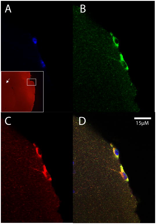
Fig. 5.
Exogenous macrophages from a CD45.1-expressing donor mouse in the superficial neocortex of a CD45.2-expressing host mouse. The cells were exposed to Aβ-rich AD brain extract in the peritoneal cavity of donor mice, collected by lavage, and injected intravenously into the host mouse 24 hours prior to sacrifice. A) DAPI nuclear stain (blue) with low magnification inset showing the location of the cells (white box) in the superficial neocortex (arrow marks the striatum); B) CD45.1 (green); C) Aβ (red); D) merged images.

