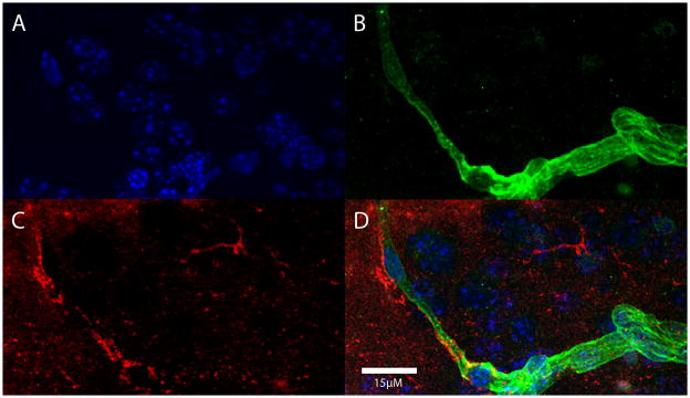
Fig. 6.
Aβ immunoreactivity (red) adjacent to a laminin-immunoreactive blood vessel (green) in the neocortex of a CD45.2-expressing host mouse 24 hours after an i.v. infusion of exogenous macrophages. In this case, the Aβ-immunoreactive cells were not demonstrably immunopositive for the donor antigen (CD45.1). A) DAPI nuclear stain (blue); B) laminin (green); C) Aβ (red); D) merged images.

