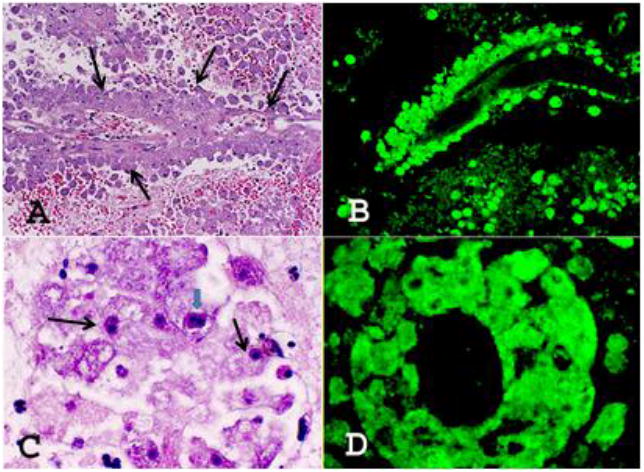
Figure 1. Liver recipient autopsy findings.

CNS sections of the liver recipient stained with H&E (A & C) and reacted with the anti-Balamuthia antibodies (B & D). A. Large numbers of Balamuthia amebae (at arrows) are seen around a blood vessel. H&E, ×200. B. A similar section reacted with the anti-Balamuthia antibodies in the immunofluorescence test. Balamuthia amebae around the blood vessel are intensely fluorescing, staining bright apple green. ×200. C. Large numbers of Balamuthia (at arrows) around a blood vessel. Note the darkly staining nucleus. A CNS section stained with H&E. One ameba is seen with double nucleolus (big arrow), × 1,000. D. A section as in B with large numbers of intensely staining apple green amebae are seen around a blood vessel, × 1,000.
