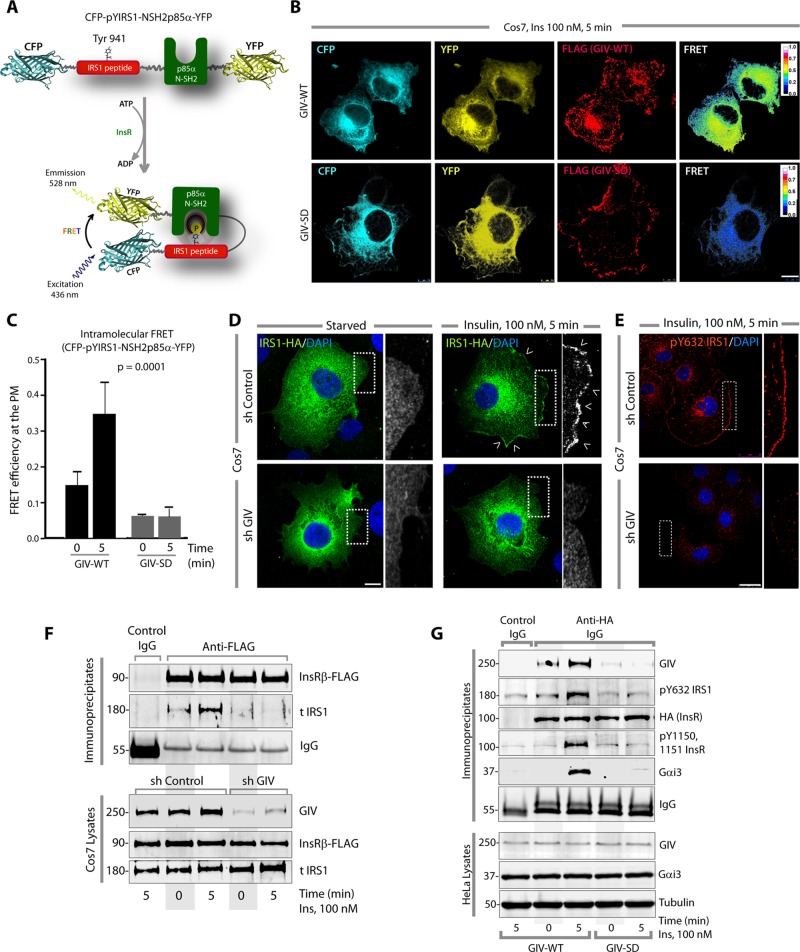
FIGURE 3:
GIV-GEF directly binds and regulates the localization and activation of IRS1. (A) Schematic for the biosensor phocus-2nes is shown. Energy transfer from CFP to YFP occurs only when Y941 is phosphorylated and the N-SH2 domain of p85α binds the phosphotyrosine ligand. (B and C) Serum-starved Cos7 cells coexpressing phocus-2nes with either GIV-WT-FLAG or GIV-SD-FLAG were stimulated with insulin, fixed, stained for FLAG (far red), and analyzed for FRET using confocal microscopy. Images panels display (from left to right, B) CFP, YFP, FLAG (GIV), and intensities of acceptor emission due to FRET in each pixel 5 min after insulin stimulation. Image panels of serum-starved (0 min) cells are shown in Supplemental Figure S3A. Bar graph (C) displays the FRET efficiency observed in GIV-WT versus GIV-SD cells at 0 and 5 min. The analysis represents five regions of interest from 4 to 6 cells/experiment (three independent experiments). Error bars = mean ± SD. (D) Serum-starved control (sh Control) or GIV-depleted (sh GIV) Cos7 cells expressing IRS1-HA were stimulated with insulin, fixed, stained for HA (green) and DAPI/DNA (blue), and analyzed by confocal microscopy. Insets show the magnification of the boxed regions. Scale bar: 10 μm. Arrowheads denote PM. (E) Serum-starved control (sh Control) or GIV-depleted (sh GIV) Cos7 cells were stimulated with insulin, fixed, stained for endogenous pY632-IRS1 (red) and DAPI/DNA (blue), and analyzed by confocal microscopy. Insets show the magnification of the boxed regions. Scale bar: 10 μm. (F) Immunoprecipitation was carried out on lysates of starved or insulin-stimulated control (sh Control) or GIV-depleted (sh GIV) Cos7 cells expressing InsRβ-FLAG. Bound immune complexes were analyzed for IRS1, InsRβ (FLAG), and IgG by IB. IRS1 coimmunoprecipitated with InsRβ in control cells but not in GIV-depleted cells. (G) GIV-depleted HeLa cells stably expressing GIV-WT or GIV-SD were transiently transfected with InsR-HA, starved, and stimulated with 100 nM insulin for 5 min before lysis. InsR and receptor-bound complexes were immunoprecipitated by incubating equal aliquots of lysates with anti-HA mAb or contro IgG, followed by protein G beads. Immune complexes were analyzed for GIV, InsR (HA), ligand-activated InsR (pY1150, 1151 InsR), pY632 IRS1, and Gαi3 by IB. Equal loading of lysates was confirmed by analyzing GIV, Gαi3, and tubulin by IB. Maximal autophosphorylation of InsR and recruitment of GIV, IRS1, and Gαi3 to the receptor was observed in cells expressing GIV-WT exclusively after insulin stimulation, but not in cells expressing GIV-SD.

