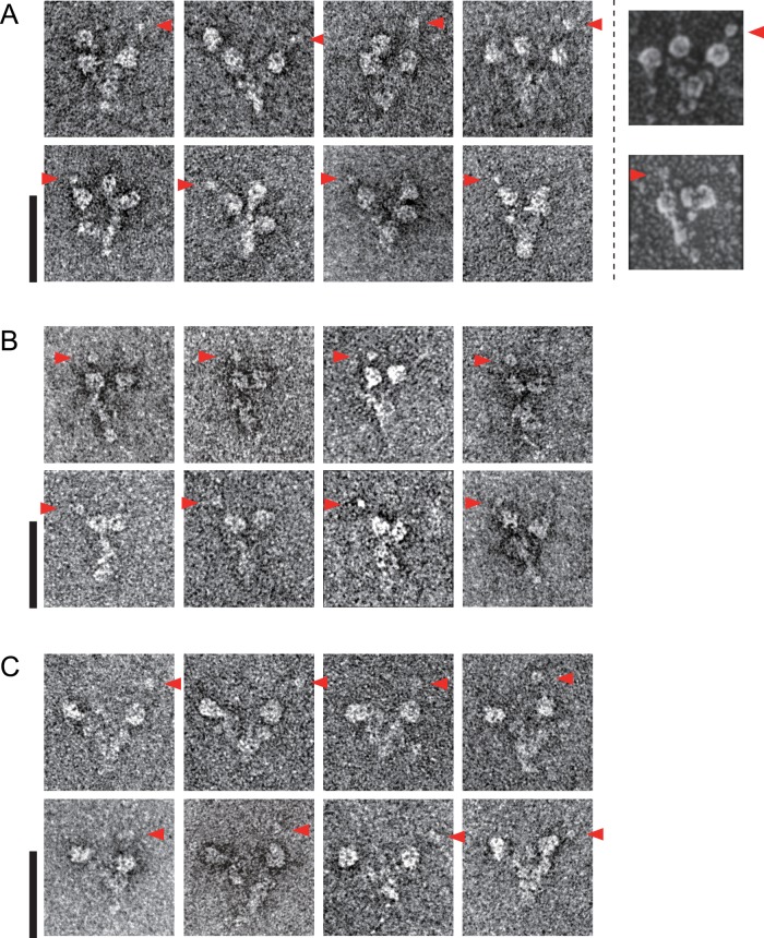
FIGURE 2:
Negatively stained EM images of Chlamydomonas OAD complex. (A) The wild- type (three-headed) Chlamydomonas OAD complex. Previous rotary shadowing EM images are shown in the rightmost column for comparison. (The rotary shadowed EM images were reproduced with permission from Goodenough and Heuser, 1984; copyright Elsevier.) (B) The oda11 (βγ two-headed) Chlamydomonas OAD complex. (C) The oda4-s7 (αγ two-headed) Chlamydomonas OAD complex. Large stalk tips are indicated by red arrowheads. Bars, 50 nm.

