. 2015 Nov 17;4:e09395. doi: 10.7554/eLife.09395

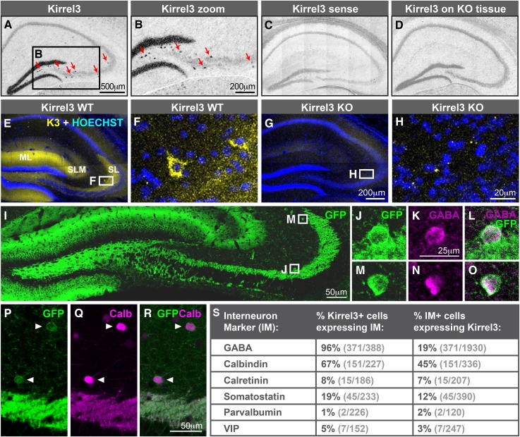
Figure 2. Hippocampal DG and GABA neurons express Kirrel3.

(AD) In situ hybridizations for Kirrel3 mRNA on adult P60–P70 hippocampal sections from WT (AC) and KO (D) mice. A negative control sense probe on WT tissue is shown in C. Red arrows in boxed region of A point to scattered Kirrel3-expressing cells shown magnified in (B). (EH) Hippocampal sections from Kirrel3 WT (E, F) and KO (G, H) mice were immunostained with anti-Kirrel3 antibodies (yellow) and Hoechst (blue). F and H are magnified images of boxed regions in E and G. (IO) P14 Kirrel3 KO mice with farnesylated GFP inserted in the Kirrel3 locus were immunostained with anti-GFP antibodies to identify Kirrel3-expressing cells (green). Dentate granule (DG) dendrites and their mossy fiber (MF) axons are brightly labeled (I) as well as GABA-expressing cells (magenta) in area CA3. (PR) P14 Kirrel3 KO mice were immunostained for GFP (green) and calbindin (Calb, magenta). (S) Analysis of Kirrel3-positive cells in P14 Kirrel3 KO mice co-expressing interneuron markers. Abbreviations: wild-type, WT; knockout, KO; molecular layer, ML; stratum lucidum, SL. Stratum lacunosum-moleculare, SLM. Images in AD, E, G, and I are tiled.

DOI: http://dx.doi.org/10.7554/eLife.09395.006

Figure 2.

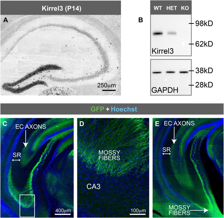
Figure 2—figure supplement 1. Kirrel3 is not expressed by CA3 neurons.

Figure 2—figure supplement 1.

(A) In situ hybridization for Kirrel3 mRNA at P14 shows the same expression pattern as the adult brain shown in Figure 2A. (B) Western blot of hippocampal lysates from adult Kirrel3 wild-type (WT), heterozygous (HET), and knockout (KO) mice indicates Kirrel3 protein is absent in knockout mice as expected. (CE) P25 Kirrel3 KO mice with farnesylated GFP inserted in the Kirrel3 locus were immunostained with anti-GFP antibodies to identify Kirrel3-expressing cells. Shown here are tiled confocal images of coronal sections through more ventral hippocampal sections compared to Figure 2E. Boxed region in C is magnified in D. Note that GFP is not present in CA3 neuron cell bodies or their axons that reside in the stratum radiatum (SR) layer. Thus, throughout the hippocampus, Kirrel3 is not expressed at detectable levels in CA3 neurons. EC; entorhinal cortex. Images A, C, and E are tiled.
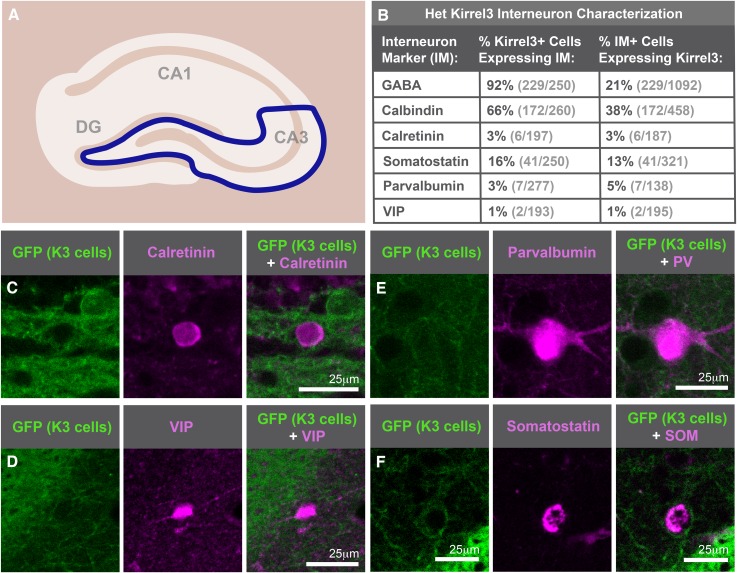
Figure 2—figure supplement 2. Kirrel3 is expressed by mainly calbindin-positive GABA neurons.

Figure 2—figure supplement 2.

(A) Diagram indicating hippocampal region imaged and analyzed is represented inside the blue line. (B) Interneuron analysis of animals heterozygous for Kirrel3 shows little difference from knockout analysis shown in Figure 2S. (CF) Sample images of interneuron staining to show each interneuron antibody worked for IHC despite having little overlap with Kirrel3. Mice are immunostained for GFP (green) to mark K3-expressing neurons and interneuron markers (magenta) in tissue.