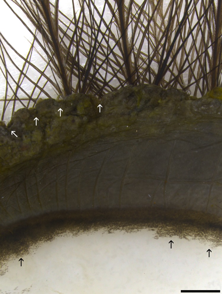
Fig 1. Marginal region of superior eyelid.
The outer marginal edge has a scalloped appearance (delineated by tips of white arrows). Approximately the outer 2/3 of the marginal zone has heavy melanization before it makes a gradual transition to the melanin-free epithelia (black arrows). Hair-like filoplume feathers with few barbs are seen arising from the epidermal side of eyelid along the marginal edge. Bar = 1000 μm.

